SAVESPACE Software Tool
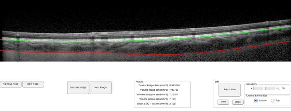
The Statistics Assistant, Volume Enumerating, Segment and Position Automatically of Choroid Edges (SAVE SPACE) Tool is a matlab GUI (Graphical User Interface) for automatically detecting the edges of the choroid layer of an OCT (Optical Coherence Tomography) scan using the Heidelberg Engineering Spectralis camera and Eye Explorer software. The SAVE SPACE tool parses XML data exported from Eye Explorer, automatically detects choroid edges of scans, allows a user to manually edit these lines for visual confirmation, and saves volumetric data of scans of all poses to facilitate statistical analysis.

The GUI was designed for Astronaut Dr. Jay C. Buckey Jr. in the Center for Spaceflight Research at the Dartmouth-Hitchcock Medical Center. I was tasked with automatically detecting choroid segments from OCT images, and provide the ability to manually change choroid lines for slight edits. In addition to noise reduction through smoothing filters and fspecial Gaussian filters, I implemented statistical classification techniques to determine choroid edges (commonly used for MRI images). Each time an eye is studied, the OCT camera takes 25 scans. Subjects' eyes were studied during multiple poses (of 25 scans each), and multiple subjects were studied. Manually segmenting all these scans is a time consuming processing that significantly slows down research output.
With statistical classification, only a handful of scans are manually segmented by "expert" segmenters (researchers who are familiar with manually segmenting images). These manually segmented images are then used to train my GUI to determine what a "good" segmentation is. Models of intensity properties of various segments (above and below) are constructed from manually segmented data. Each pixel of the image to be automatically segmented is compared against the models and assigned a most probable or likely category, assuming image intensities are Gaussian.
The final program takes in structure and images exported from the OCT software, automatically segments all scans and calculates choroid volume, allows a user to cycle between 25 scans within a pose and between poses, provides the ability to edit (with undo, redo, remove functions, as well as ability to change parameters of a spline fill), and has the capability to populate a matrix of appropriate subject data for easy statistical analysis with whatever prefered statistical analysis software.
It should be stated that the initial portion of this project was influenced by the work previously done by David Alonso-Caneiro from Queensland University of Technology. His white paper describing another method for segmenting images (not calculating volume) can be found here.

